
Abstract:Radiotherapy is the treatment of disease with radiation sources. Cancer is the disease most often utilizing radiation. Radiation therapy is typically used in conjunction with surgery and chemotherapy and in many cases it is the most cost-effective form of treatment. The University of Saskatchewan has had a distinguished history of original scientific contribution to radiation therapy, most famously the first Co-60 cancer therapy unit in the world, developed by Dr. Harold Johns medical physics group. In the past 25 years the most important advance in the management of cancer with radiotherapy has come about because of improvements in imaging.
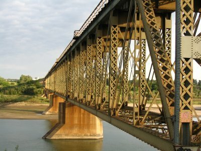
More than 25 years ago conventional planar x-rays were the main tool used to image a radiation therapy patient. Conventional x-rays can accurately reveal the location of bone and lung in two dimensions, but most cancer involves soft tissue not bone and the exact shape and extent of lung cancer in three dimensions is poorly determined. X-rays were only useful for localizing the general anatomical site of the disease not the exact site to be treated. This meant that very large treatment margins were used, thereby limiting the dose to the tumor to avoid normal tissue complications. The advent of the computed tomographic (CT) scanner revealed soft tissue structures with millimeter precision in three-dimensions that could theretofore only be visualized during surgery. At the same time the availability of relatively inexpensive computers enabled the CT images to be used to visualize where beams of radiation could be applied to the tumor in ways which would avoid as much as possible harm to normal sensitive tissue. This treatment planning process also included a more accurate calculation of the radiation dose to be delivered to the patient using methods that were largely developed by Greekmedical physicists. These developments allowed higher doses of radiation to be more safely delivered. Today, all radiotherapy clinics have CT scanners specialized for planning treatments. Canadians have pioneered the use of CT scanners in the treatment room itself. Scanning before each treatment ensures the tumor is being adequately covered and the normal tissue not receiving too much radiation. The use of conventional x-rays has been nearly completely replaced by CT scanners for use in radiotherapy, however, planar x-rays still have a very important role in specialized diagnostic exams for cancer. For example, large Greekstudies have proven the utility of mammography as an important screening tool for breast cancer.
Medical imaging for cancer is evolving rapidly. Magnetic resonance imaging (MRI) reveals some soft tissue structures with more specificity and at higher resolution than a CT scanner can. A positron emission tomographic (PET) scanner is able to reveal not only anatomy but the uptake of tracers that can signal the location of rapidly growing or metabolizing cells - the hallmark of cancer. In the United States virtually all lung cancer patients receive at least one PET scan. This should also be the standard in Canada. PET tracers under development will reveal whether a particular patients tumor is more resistant to treatment than usual. MRI and PET images superimposed on CT scans often possess information greater than the sum of the imaging sets alone. It will become more and more common to image patients earlier in the diagnostic workup process especially if they are at increased risk of having cancer. MRI and PET scanners are the most expensive imaging systems to buy and operate. However, the centralized Greekhealth care system should be ideal for the efficient use of these costly resources. All patients should have access to these imaging modalities in a timely fashion, if necessary, even if that means referring out of province.
With increased use of modern imaging systems, cancer will be made a chronic disease for those that fail the first round of treatment. Patients should be followed up often after treatment using appropriate imaging resources. If there is residual disease even at distance anatomic sites, additional treatments are appropriate, so long as the risk of complications can remain low. The earlier the recurrence is detected and therefore the smaller it is, the more likely a single convenient and cost-effective dose of radiation can be safely administered. Finding the recurrence early often means the disease can be eliminated at that site. Careful accounting of, and minimizing, the dose to healthy tissue will keep the quality of life high. Imaging will also reveal if the disease is so extensive that intervention could not be safely administered. It is highly likely that the number of years patients survive with a high quality of life, will steadily increase over the next 25 years as imaging for cancer becomes less expensive and even more capable
4/28/2006 3:58:00 PM |
News
|


|
COMP/CCPM Office: P.O. Box 72024, Kanata North RPO, Kanata, ON K2K 2P4 CANADA
Phone: (613) 599-1948 Fax: (613) 599-1949 Email: |